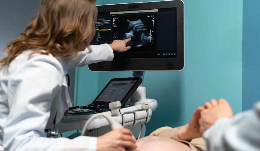
Introduction for Fetal ECHO Scan
A Fetal Cardiac Scan in Thoraipakkam is a specialized ultrasound performed to evaluate the structure and function of your baby’s heart in the womb. Also known as a fetal echocardiogram, this scan allows early detection of congenital heart defects, enabling timely planning for care immediately after birth if needed. At MOM Fetal Scans, I perform this scan using advanced imaging technology, ensuring precise results and complete reassurance for parents. Many mothers search for fetal echoes Thoraipakkam or fetal heart scan Thoraipakkam to find expert-led cardiac assessments during pregnancy.
This scan goes beyond routine ultrasounds by giving a detailed view of heart chambers, valves, major vessels, and blood flow patterns. Observing the heart carefully helps identify structural or functional issues early, giving parents confidence and a clear understanding of their baby’s health. My goal is to provide both medical clarity and emotional reassurance throughout the procedure.
What Is a Fetal ECHO Scan?
A fetal cardiac scan, or fetal echocardiogram, is an advanced ultrasound that examines the anatomy and function of the baby’s heart. It evaluates the size and structure of the heart chambers, valves, outflow tracts, and major blood vessels, as well as heart rhythm and blood flow. Performing this scan ensures that any congenital heart defects are identified early, allowing timely interventions if required. Many expectant parents in Thoraipakkam seek this as a fetal echo test for pregnancy to ensure accurate cardiac evaluation.
In addition to anatomical assessment, the fetal cardiac scan monitors how effectively the heart pumps blood and whether circulation is proper. At MOM Fetal Scans, findings are documented in a structured format to guide future follow-up or specialized care if needed. The scan is particularly important for mothers with diabetes, abnormal ultrasound findings, family history of heart defects, or multiple pregnancies. Early knowledge empowers parents with reassurance and actionable information.
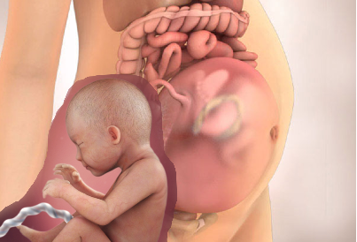
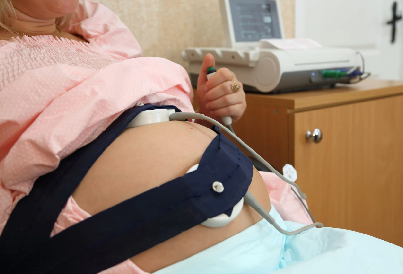
Key Elements of This Scan
- Chamber and valve evaluation – Checks heart structure and function
- Major vessels and outflow tracts – Detects anomalies
- Blood flow assessment – Ensures proper circulation
- Heart rhythm monitoring – Identifies irregularities early
- Documentation for follow-up – Supports planning and management
When Should a Fetal ECHO Scan Be Done?
A detailed fetal heart scan is usually performed between 18 and 22 weeks of pregnancy. This timeframe allows optimal clarity for evaluating cardiac structures, blood flow, and heart rhythm. In high-risk pregnancies, earlier or repeat scans may be recommended to monitor development closely. Parents searching for fetal echocardiography test near me often prefer this period for the most accurate assessment.
Performing the scan too early may limit visibility of some structures, while scanning beyond 22 weeks can reduce image clarity due to fetal positioning and growth. Scheduling within this window allows precise assessment of heart chambers, valves, vessels, and overall function, ensuring parents and doctors have clear insights into the baby’s heart health.
Why Timing Matters
- Optimal visualization of heart structures
- Accurate assessment of valves and vessels
- Reliable blood flow evaluation
- Heart rhythm monitoring possible
- Early detection of congenital defects
Who Should Get a Fetal ECHO Scan?
All pregnant women may benefit from a fetal cardiac scan, but it is especially important for mothers at higher risk of congenital heart defects. This includes mothers with diabetes, hypertension, autoimmune disorders, previous pregnancies with heart anomalies, or a family history of congenital heart conditions. Multiple pregnancies also benefit from detailed cardiac evaluation for each baby.
Parents often search for a fetal echo test for pregnant ladies in Thoraipakkam to gain early reassurance. A fetal cardiac scan provides early detection of potential heart issues, guiding prenatal care, delivery planning, and postnatal interventions if required. I ensure that every mother receives personalized attention, clear explanations, and compassionate support throughout the procedure.
Recommended For
- All pregnancies (routine or reassurance scan)
- High-risk mothers
- Maternal diabetes or hypertension
- Family history of heart defects
- Multiple pregnancies or IVF cases
How Is the Fetal ECHO Scan Performed?
The scan is a non-invasive ultrasound procedure performed over the mother’s abdomen. Warm gel is applied, and a high-resolution ultrasound probe captures images of the heart from multiple angles. At MOM Fetal Scans, we use advanced imaging technology to provide detailed visualization of heart chambers, valves, outflow tracts, and vessels. The procedure typically takes 20–40 minutes, depending on the baby’s position and activity.
During the scan, I explain the findings in real-time, showing how blood flows through the heart, the heartbeat, and the structural anatomy. Parents can see their baby’s heart functioning and understand the health status. All findings are carefully documented to guide follow-up or any further specialized evaluation if needed.
During the Scan You Will Experience
- Comfortable, non-invasive procedure
- High-resolution visualization of organs
- Real-time explanation of findings
- Observation of fetal heart movements
- Accurate documentation for follow-up
What Does the Scan Check in Detail?
The fetal cardiac scan evaluates the structure and function of the heart. The heart chambers, valves, major vessels, and outflow tracts are examined for normal size, alignment, and connections. Blood flow through the heart and vessels is checked using Doppler technology to detect abnormalities in circulation or rhythm. The heart rate is monitored, and structural anomalies such as septal defects or valve malformations are identified if present.
This detailed assessment provides reassurance to parents and enables early planning if intervention is needed after birth. By recording all findings, the scan ensures proper documentation and tracking of fetal heart development. Parents searching for fetal heart scan Thoraipakkam or fetal echoes Thoraipakkam choose this scan for its thorough evaluation and reliability.
Structures We Examine
- Heart chambers and valves
- Major vessels and outflow tracts
- Blood flow and circulation
- Heart rhythm and rate
- Overall cardiac function and structure
How to Prepare for the Scan?
No special preparation or fasting is required for a fetal cardiac scan. You may be asked to have a moderately full bladder to improve imaging clarity. Wear loose and comfortable clothing for easy access to the abdomen. Bring any previous scans or reports that may help compare growth and development.
Staying calm and relaxed will help the scan go smoothly. I ensure that every mother feels supported and informed, explaining each step and observation in a compassionate manner. Parents often find the real-time explanation reassuring, especially when understanding the details of their baby’s heart.
Preparation Tips
- No fasting required
- Moderately full bladder may help imaging
- Wear comfortable clothing
- Bring previous scan reports
- Stay relaxed for a smoother scan experience
What Happens After the Scan?
After the scan, you will receive a detailed report outlining all findings, including heart chambers, valves, blood flow, and any detected anomalies. If results are normal, routine prenatal care can continue with confidence. If any concerns are identified, I will guide you through follow-up scans or specialist referrals for optimal management. Early detection allows timely interventions for the best possible outcomes.
Parents often leave this scan feeling reassured after seeing their baby’s heart in motion. My focus is to provide clarity, guidance, and support, ensuring you understand the results and next steps completely.
After-Scan Guidance
- Detailed report with findings
- Clear explanation of results
- Follow-up scans if required
- Routine care if normal
- Supportive counseling for parents
Conclusion
The Fetal ECHO Scan in Thoraipakkam is a vital step in your pregnancy, offering detailed insight into your baby’s heart development. At MOM Fetal Scans, I combine advanced imaging technology with compassionate care to ensure mothers are fully informed and reassured. This scan is crucial for detecting potential congenital heart defects early, supporting a safe and healthy pregnancy journey.
Book your Fetal Cardiac Scan at MOM Fetal Scans Thoraipakkam today for expert assessment, precise results, and complete reassurance for you and your baby.

