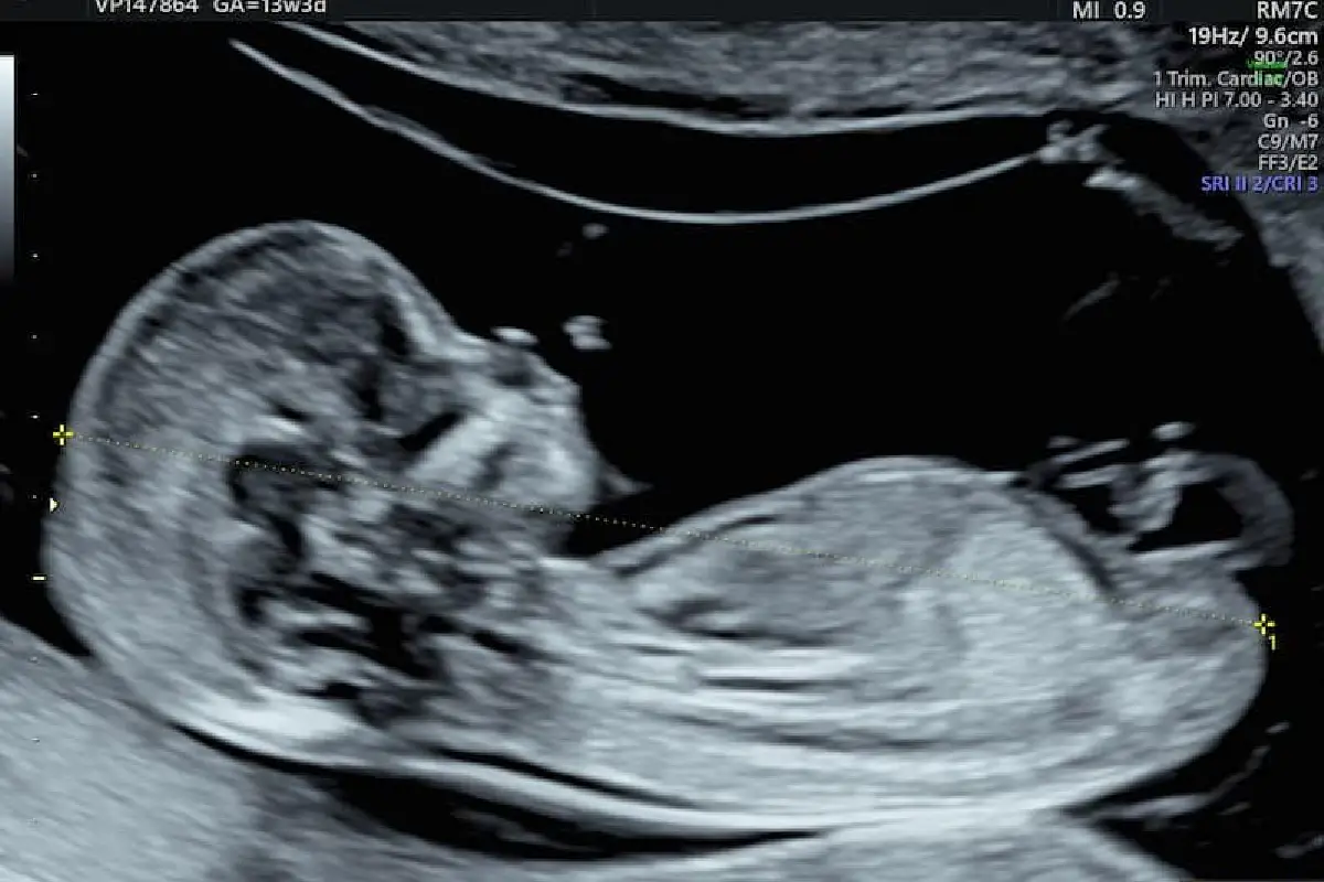
Introduction for Early Anatomy & Reassurance Scan
As a Fetal Medicine Specialist at MOM Fetal Scans, I understand how vital the second trimester is for every expectant mother. Between 14–19 weeks, your baby undergoes rapid development, making this period ideal for a more detailed fetal assessment. The Early Anatomy & Reassurance Scan is designed to evaluate your baby’s growth, movement, organ formation, and overall wellbeing. Many mothers search for terms like anatomy scan Thoraipakkam, OMR, fetal anatomy scan near me, anatomy ultrasound pregnancy, or reassurance scan, and this service page will guide you through everything clearly and supportively
This scan is especially helpful for mothers who want early reassurance before the standard anomaly scan at 18–22 weeks. It offers a confident, structured overview of your baby’s major organs, limbs, spine, heartbeat, and growth patterns. At MOM Fetal Scans Thoraipakkam, OMR, I ensure that every scan is performed safely, compassionately, and with 100% accuracy using advanced ultrasound technology. This page will walk you through what the scan is, when to do it, why it matters, how to prepare, and what to expect afterward.
What Is the Early Anatomy & Reassurance Scan?
The Early Anatomy & Reassurance Scan is a focused evaluation performed during 14–19 weeks of pregnancy to examine your baby’s developing organs, bones, brain structures, and overall growth. Unlike a basic ultrasound, this is a more structured assessment that looks at important fetal features earlier than the detailed anomaly scan. Many mothers who search for fetal anatomy scan Thoraipakkam, OMR or anatomy ultrasound pregnancy prefer this scan for early confidence and clarity.
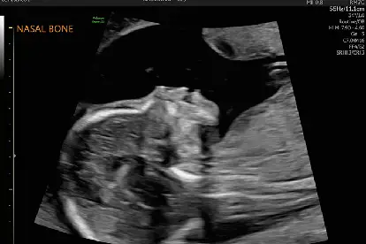
Though it is not a replacement for the full anomaly scan at 18–22 weeks, this scan provides an early look at the baby’s anatomy. I carefully evaluate the skull, brain, spine, stomach, kidneys, limbs, heart chamber positions, and placenta. This helps detect major developmental concerns early and gives parents reassurance about fetal wellbeing. Every observation is explained in simple and supportive language, helping you understand each part of your baby’s development.
Why This Scan Matters
- Early assessment of fetal anatomy
- Checks organ formation and development
- Provides early reassurance for mothers
- Helps detect major abnormalities earlier
- Assesses placenta, fluid, and fetal growth
When Should You Get This Scan?
This scan is ideally performed between 14–19 weeks. Before 14 weeks, many organs are still too small to evaluate accurately. After 19 weeks, the scan becomes part of the routine anomaly evaluation. Therefore, this early window gives the perfect opportunity to check organ development while still providing enough time for follow-up tests if needed.
Many first-time mothers opt for this scan around 16 weeks, when the baby’s structures are clearer. Mothers who previously experienced pregnancy loss or complications may choose this reassurance scan earlier within the window. Whether you visit for reassurance or anatomical clarity, this timing ensures you receive accurate and meaningful information about your baby’s progress.
Why the Timing Is Important
- Ensures optimal visibility of organs
- Helps plan follow-up tests in time
- Provides reassurance early in the second trimester
- Avoids overlapping with the detailed anomaly scan window
- Ensures baby’s movements assist in clear imaging
Who Should Get an Early Anatomy & Reassurance Scan?
This scan is recommended for all mothers who want early clarity about fetal development. While every pregnancy can benefit from this early anatomy ultrasound, certain mothers need it especially. Women with previous miscarriages, IVF pregnancies, diabetes, thyroid issues, bleeding, or anxiety about fetal development often choose this scan for peace of mind.
It is also advisable for mothers who have concerns about fetal movement, abdominal pain, or growth patterns. Families who simply want early visualization of their baby’s organs also benefit greatly. Whatever your reason, this scan ensures that you enter the second trimester with confidence and reassurance.
Best Suited For
- First-time mothers
- IVF or ICSI pregnancies
- Mothers with previous complications
- Women with medical risk factors
- Anyone seeking early reassurance
How Is the Scan Performed?
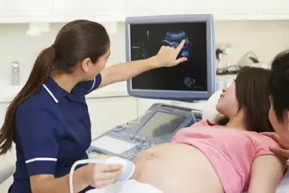
This scan is done through an abdominal anatomy ultrasound. A warm gel is applied to your abdomen, and the ultrasound probe helps visualise the baby in detail. The process usually lasts 10–20 minutes, depending on your baby’s position and activity. As the baby moves more during this stage, we often get beautiful views of the face, limbs, heartbeat, and spine.
During the scan, I examine the early brain structures, spine alignment, stomach bubble, bladder filling, limb growth, and heart positioning. You will see your baby moving, stretching, and even kicking — making this a deeply emotional and memorable experience. The entire procedure is safe, painless, and comfortable.
What You Will Experience
- Comfortable abdominal ultrasound
- Clear view of organs and limbs
- Evaluation of heartbeat & growth
- Early screening for major conditions
- A memorable bonding moment with your baby
What Does This Scan Detect?
The Early Anatomy & Reassurance Scan gives an early look at major organ development, but it is not a complete anomaly scan. It is designed to detect concerns such as abnormal skull shape, abdominal wall defects, major limb abnormalities, absence of stomach bubble, or early spinal concerns. It also checks placental position, amniotic fluid volume, fetal movement, and growth.
While it cannot detect all conditions, it provides essential information about your baby’s wellbeing much earlier than the detailed anomaly scan. This helps plan next steps early and reduces anxiety for expecting mothers.
Detects Early Indicators Of
- Major structural abnormalities
- Brain and skull development concerns
- Limb formation issues
- Abdominal or spinal defects
- Placental and fluid-related issues
How to Prepare for This Scan?
Preparation for this scan is simple. You do not need to fast. A moderately full bladder may help improve clarity, especially earlier in the 14–16 week window. Wearing comfortable clothing helps you lie down easily during the scan. Bringing previous ultrasound reports or blood test results allows me to compare progress accurately.
Emotionally, preparing with a calm and positive mindset helps you enjoy the experience fully. Many mothers feel reassured just by seeing their baby moving actively — preparation is minimal but the experience is meaningful.
Preparation Checklist
- Keep a moderately full bladder
- Wear loose, comfortable clothes
- Bring previous medical records
- Arrive a few minutes early
- Stay relaxed for better imaging
Are There Risks or Limitations?
The Early Anatomy & Reassurance Scan is completely safe, using ultrasound waves with no radiation. There are no risks to you or your baby. However, like all ultrasound screenings, it has limitations. Some organs are still developing, so finer details of the heart, face, or brain may not be visible until 18–22 weeks.
Baby’s position can sometimes limit what we can see clearly. If certain views are unclear, I may ask you to walk, change position, or return after a short break. Although this scan gives valuable early information, it is still not a replacement for the detailed anomaly scan.
Limitations to Know
- Not as detailed as an anomaly scan
- Some organs still developing
- Baby’s position may restrict views
- May require follow-up scans
- Does not detect every condition
What Happens After the Scan?
Once the scan is complete, I review the baby’s organ development, growth parameters, placenta, fluid, heartbeat, and movements. I explain the findings clearly so you understand the significance of each observation. If everything looks normal, this scan provides strong reassurance for the upcoming second trimester.
If any concerns are found, I will guide you through next steps such as repeat scans, anomaly scan scheduling, or genetic counseling if required. My goal is to ensure that every mother feels supported and well-informed — never anxious or confused
After-Scan Outcomes
- Detailed report provided
- Explanation of every finding
- Guidance on follow-up scans
- Early detection of concerns
- Emotional reassurance for parents
Conclusion
The Early Anatomy & Reassurance Scan (14–19 weeks) is an essential milestone in your pregnancy journey. It offers a structured look at your baby’s early anatomy, providing comfort, clarity, and confidence as you enter the second trimester. Whether you’re searching for anatomy scan Thoraipakkam, OMR, fetal anatomy scan near me, anatomy ultrasound, or reassurance scan, this evaluation ensures that you receive expert attention and meaningful insights.
At MOM Fetal Scans in Thoraipakkam, OMR I, Dr. Subhashini Ravi, ensure that every scan is performed with accuracy, empathy, and clear communication. My priority is the wellbeing of both mother and baby — and this early anatomy scan provides a strong foundation for healthy pregnancy care.
Book your Early Anatomy & Reassurance Scan at MOM Fetal Scans Thoraipakkam, OMR today and step into your second trimester with confidence, clarity, and expert fetal care.
Frequently asked question
No. An early anatomy scan gives a preliminary assessment of major organs, while the detailed anomaly scan (18–22 weeks) provides a full, in-depth evaluation of the baby’s development.The two scans complement each other but are not interchangeable
Yes. A reassurance scan can confirm twin or multiple pregnancies, show the number of sacs, placentas, and check heartbeat and early growth.It helps determine chorionicity and amnionicity early.
No fasting is needed. You may only be asked to have a moderately full bladder for clearer images in early pregnancy.This makes the scan simple and convenient for mothers.
Some major heart abnormalities can be seen early, but a detailed fetal echo or the anomaly scan at 18–22 weeks is more accurate for detecting cardiac issues.Early views provide preliminary clues but not the full picture.
Usually 10–15 minutes, depending on the baby’s position and the level of detail needed.Some scans may take slightly longer if the baby is very active.

